Course Learning Outcomes
• Understand the factors that influence healthy fascia and relate them to clients and participants
• Understand how fascia connects everything in the body
• Explain the different roles fascia plays in the body
• Apply a wholistic approach to working with clients and participants
• Understand basic movement strategies and SMR techniques to enhance fitness performance and quality of life
MODULE 1
History
2500-3000 BCE- Medical writers were aware of fascia through the surgeries being performed
600 BCE—Fascia identified as connective tissue
1600’s–The word fascia enters English medical writing
1700’s–1st PubMed article to contain the word fascia is published. Refers to fascia as tissue that separates muscle and supports movement
1800’s–Fascia described generically as undifferentiated tissue that covers muscle and is found under the skin. Also described as packing material between other more specialized tissues
1900’s–Knowledge and interest in fascia grows significantly due to development of powerful microscopes and formaldehyde
2000’s–Understanding of the complexity of fascia increases with awareness that it takes in sensory input and is involved in producing skeletal muscle movements (stores mechanical energy
Functionality of fascia
Intakes sensory input
Stores mechanical energy
Multiple layers or areas
Influences all areas of the body
KEY TERMS
CONNECTIVE TISSUE-
General term referring to tissues that protect and support the overall structure of tissues and organs. Helps connect tissue to other tissue. The 4 types of connective tissues are blood, bone, cartilage and connective tissue proper. Fascia is a subtype of connective tissue
DEEP FASCIA-
The fibrous layer of connective tissue found beneath superficial fascia. There are two categories; aponeurotic fascia and epimysial fascia. Contains no fat
MYOFASCIAL CHAINS/TRAINS/SLINGS-
Pathways of muscle and fasciae that run through the entire body. Members work together in the chain to create stability and movement, and transfer tension/load from one region of the body to another close-by or far-removed regions
SUPERFICIAL FASCIA-
Fascia found directly under the skin and attached to it via Retinacula Cutis Superficialis Fibers. Contains fat
MODULE 2
Fascia’s:
1.cellular components
2.types (deep, superficial, visceral, parietal)
3.key roles in move and sensation (mechanical-non-mech)
4.factors to optimal and non-optimal fascia
detailed breakdown: AI-gen
1. Superficial Fascia:
Also known as subcutaneous tissue, Located directly under the skin, Contains loose connective tissue and adipose tissue, and Varies in character across different body regions.
2. Deep Fascia:
Surrounds muscles, bones, nerves, and blood vessels.
More fibrous and resilient than superficial fascia.
Forms fascial compartments, separating groups of muscles.
Can be further classified as axial (around the spine) or appendicular (around limbs).
3. Visceral Fascia:
Surrounds internal organs and supports them within body cavities.
Can also refer to the fascia of the brain, meningeal fascia.
4. Parietal Fascia:
Lies on the inside of body cavities and lines the walls.
Found in the pelvis and other areas.
Definition of Fascia:
-Fascia is a 3-D continuum of solf collagen-containing connective tissues found throughout the body.
It incorporates:
-adapost tissue=fat,
-neurovascular sheet =blood vessels and nerves,
- periosteum=bone,
-muscle tissue
Fascia anatomically separates systems. However these systems work together with support of fascia.
It is a type of connective tissue. This category of tissue covers a vast majority of what makes up our body.
Fascia exists outside of the standard anatomical bc there are no fixed start and end points.
Four Types of Tissue in the Bodye
Connective Tissue
Tissue that helps connect things together along with contributing to form and structure of the body. Eg. Myofascia: considered the scaffolding for muscles. This term refers to the fascia specifically for muscles
Epithelial
Barrier= Type of tissue that forms the outer covering of all internal and external surfaces of the body (Eg. body cavities, hollow organs, glands, arteries, veins, GI tract). Our skin is considered epithelium or epithelial tissue.Eg. skin
Muscle
Contractile soft tissue found in animals and humans. Muscle cells are comprised of contractile proteins called myosin and actin. Contraction and elongation of the proteins result in a change in the length and shape of the cell.
Eg. skeletal, cardiac, smooth
Nerve Tissue (Nerves)
Communication lines between one part of the body to another. Elongated nerves cells that connect the body to the spinal cord and brain, (Eg. Central Nervous System), which is the control centre of the body. Information passes by electrical nerve impulses from neuron to neuron until it reaches its target location.Eg. neuronsplete.


Cells- build fibers and secrete ground substance
Fibroblasts - main cellular component
Adipocytes- fat cells
Chondrocytes- cartilage cells
Osteocytes- bone cells
Macrophages and mast cells- Immune cells. White blood cells
Fibers - link everything, physical connection
Ground sub - extracellular matrix, fluid that keeps components healthy and functioning optimally


What is Fascia?
• Fat, blood vessels, nerves, bone, muscle tissue
• Anatomically separate systems
• Work together with the support of fascia
• Ideal to consider anatomical and functional models of fascia

Fascial Anatomy

Epidermis and Dermis:
Tissue that makes up the skin
Superficial Fascia:
Loose connective tissue located directly underneath the skin. Contains fat
Deep Fascia:
Dense, fibrous tissue that contains no fat. Two types: aponeurotic and epimysium
Epimysium:
The most superficial layer of fascia that goes around muscle. Helps prevent friction between adjacent muscles
Muscle:
Specialized tissue made up of thin, elongated cells. Contract to produce voluntary or involuntary movement
Superficial Retinacula Cutis Fibers :
Act as skin ligaments; fibers that connect the dermis layer of skin with the superficial fascia
Adipose Tissue:
Tissue made up of primarily triglycerides
Deep Retinacula Cutis Fibers:
Fibers that connect superficial fascia to deep fascia
Hyaluronic Acid:
GAG molecule found in ground substance/ECM
What is Fascia
Endows the body with functional structure
Enables the body systems to operate in an integrated manner
Surrounds, interweaves and penetrates all organs, muscles, bones and nerve fibers for functional purposes
Subcategories of Connective Tissue
Bone Mineralized connective tissue that creates the hard structure of the human body. Contributes to locomotion, support, protection of soft tissues and organs and storage system for minerals (calcium, phosphorus)
Ligament A dense fibrous connective tissue band connecting bone to bone, providing structure stability or integrity to joints
Tendon A strong, dense fibrous connective tissue band that attaches muscles to bones. Responsible for transmitting mechanical forces created in the muscles to the bones, resulting in movement. Made of numerous collagen fibers
Tensegrity–Describes a 3-dimensional structure consisting of members or tissues under tension that are contiguous with members or tissues under compression. The physical structure is held in place by tensile and compression forces acting at the same time. The term, when applied to a biological framework, is referred to as biotensegrity.
Biotensegrity–Describes biological structures like muscles, bone, ligaments, tendons and fascia combined to produce a strong connected bond via tensioned and compressed components.
Aponeurotic- is a type of deep fascia, a connective tissue layer that envelops muscles and helps them attach to bones
KEEPING FASCIA HEALTHY
Movement promotes flow of fluid
Promoting exercise, movement and healthy lifestyle = healthy fascia
HEALTHY FASCIA vs, DENSIFICATION
Lack of movement, dehydration, injury = densification
Addition of more fibers
Leads to non-optimal fascial movement
WHAT RESTRICTS FASCIA?
Insufficient hydration
Excessive fitness training
Excessive physical labour
Daily emotional stress
Inability to relax
Insufficient sleep
Immobility
Poor diet
Anxiety
Restricted Fascia Leads To
-Tight Muscles
- Stiff Joints
• Restricted ROM
• Restricted General Mobility
- Decreased Strength
- Decreased Speed
• Decreased Balance
• Poor Posture
Keeping Fascia Healthy
• Hydration Levels
• Appropriate training to rest ratio
• Sleep Quality and Quantity
• Occupation
• Lifestyle/Daily Activity (outside the gym)
• Healthy Food Choices
* Stress Levels
Fascia is a connective tissue
As such, we can predict how it will act based on its components
Unique tissue that connects all musculoskeletal components
Also connects with all other body parts (organs, ligaments, joints, & bone)
These connections are termed fascial systems or lines
Healthier fascia means a healthier body
QUIZ
Fascia is a type of connective tissue
Two important factors in healthy fascia are adequate hydration and activity levels.
Three components of connective tissue are cells, fibers and ground substance .
Two types of connective tissue discussed in this module were superficial and deep fascia.
Two fiber types that make up fascia are collagen and elastin.
Deep fascia can be further subdivided into aponeurotic , epimysium, visceral and parietal fascia.
Module 3
Outcomes
-Describe the types of fascial tissue and locate examples of each
-Describe the cellular components, fibrous components and extracellular substances of fascia and their specific roles
-Explore the physical relationships and communication between fascia and other tissues and how they work together
- Uncover how fascial pathways influence movement patterns and b mechanics
- Discuss how certain pathologies are connected to fascia
the types of fascia we find in the body and how they vary. We go into detail about how their cells, fibers and substances vary and how that impacts the roles each plays in the body.


Superficial FasciaComplete.
Cutaneous Nerves
Cutaneous nerves - Sensory nerves found in fascia and other tissues which are essential for survival. Provide information from the internal and external environment back to the Central Nervous System.
Eg. mechanoreceptors, thermoreceptors, nociceptors
Retinacula cutis- Found in multiple locations, when located underneath the skin they are called Retinacula Cutis Superficialis. Skin ligaments that allow the skin and superficial fascia to slide and glide.
Superficial fascia
Loose connective tissue located directly underneath the skin, responsible for acting as a mechanical and thermal cushion. Fibers run in a randomized pattern. Contains cutaneous nerves, blood vessels and lymphatic vessels.
Blood vessels
Provide tissues with oxygen and remove waste products.
Deep Fascia
Dense, fibrous tissue that contains no fat. Two types: aponeurotic and epimysium
Lymph Channels
Collect extra fluids from cells and tissues that isn't reabsorbed
DEEP FASCIA

Two subcategories: Aponeurotic and Epimysal
No fat content (more dense, less manipulative)
Sleeves and surrounds muscles and organs

Not as manipulative as superficial
Concentrated hyaluronic acid molecules
Consists of 2-3 layers of parallel collagen fibers (different angulations) separated by a layer of loose connective tissue

Anchors superficial fascia and other tissues
Protective sheaths around nerves and blood vessels
Merges around joints and organs to provide support and strength
Distributes forces evenly across muscles ans joints during movement

Thins out to surround tendons -acts as an attachment point
Adaptable - remodels in response to mechanical stressors (exercise)
Optimizes tissue function and structural integrity

2-3 layers
Within each layer fibers are parallel
Between layers- varying angulations of 75-80 *
Load layers in each direction during exercise
Biomechanical property influence movement efficiency and performance

Fascial lines
Create continuity among muscles involves in same movement
Form basis of myofascial chains
Smooth force transmission across tissues and joints within fascial lines/chains
Transmit proprioceptive info (i,e. Length, tension, movement)
TYPES OF DEEP FASCIA
APONEUROTIC FASCIA
• Broad, flat sheets or bands
• Superficial to Muscles
• Collagen and elastic fibers provide flexibility and recoil
- site for muscle attachments (30%)
• Transmits forces generated by muscles to adjacent structures
• Stabilizes muscles and transmits forces
• Reduces friction between muscle layers
-Allows for efficient muscle contraction and movement without resistance
EPIMYSIAL FASCIA
features
• Envelopes individual muscle, provides their shape and integrity
• Protective covering
• Structural support
• Facilitates movement
• Maintains shape
functions
• Prevents overstretching
• Prevents trauma, friction, injury
• Compartmentalizes muscles for optimal function
QUIZ
Aponeurotic and Epimysial:
two types of DEEP FASCIA
Hyaluronic Acid -in DEEP FASCIA
DEEP FASCIA more dense than Superficial fascia
2-3 layer of Collagen fibers (fibers run at different angles) -DEEP FASCIA
Doesn’t store mechanical energy - SUPERFICIAL FASCIA
Can be manipulated through the skin - SUPERFICIAL FASCIA
Loose connective tissue = SUPERFICIAL FASCIA
Random fiber pattern = SUPERFICIAL FASCIA
Contains fat =SUPERFICIAL FASCIA
Cellular Structures of Fascia.
Cellular components:

Connective tissue= CELLS+FIBERS+GROUND SUBSTANCE
Cells= Fibroblasts+Adipocytes+Macrophags, Mast Cells+Chondroblasts/Chondrocytes, Osteoblasts/Osteocytes
Fibers= Collagen + Elastin
Ground substance = Glycosaminoglycan + Water + Ions

Fibroblasts:
Most important cell in connective tissue
Found everywhere
Synthesizes collagen, elastin, and ECM/ground substance

Adipocytes:
Fat cells, store and release energy
Primary fat molecule - Triglycerides
Mechanical protection and temperature regulation
Direct relationship between size and how much Triglyceride stored

Macrophages and Mast Cells:
Subgroups of White Blood Cells
Primary Immune Cells
Function as Phagocytes - destroy foreign materials, damaged cells, microorganisms, dead cells, bacteria, parasites

Chondrocytes and Osteocytes:
Chondrocytes - Cartilage cells
Osteocytes - Bone Cells
Chonoblasts and osteoblasts activate to produce cartilage and bone
More active during growth, exercise, movement

Collagen:
Provide structure, support and connection to fascia matrix
Greek word for “glue”
29 subgroups
Made from different combinations of protein to create fibers
Types I to V are most common

Collagen:
Type I - most common (90%) skin, tendons, ligament, bone. Least elastic
Type II - more elastic and softer – nose, ears, joints
Type III - Blood vessels, muscles, organs —-provides structural support.

Elastin:
Second most common fiber
Gives tissue their viscoelastic properties (low stiffness, extensibility, elastic energy, storage)

Ground substance - Water:
Essential for optimal functioning
Maintains tissue pliability and elasticity
Stores GAG molecules ans ions
Decreases with age, injury, inactivity
Less that optimal hydration increases risk of injury

Ground Substance - GAG:
Glycosaminoglycans
Large sugar molecules
Most common - hyaluronic acid

GAG and Hyaluronic Acid:
Important to synovial fluid, decrease of which contributes to osteoarthritis
Participates in tissue and wound healing
Role in slide and glide mechanism
KEY TERMS and CONCEPTS
Blood Vessels
Closed loop channels that carry blood throughout the body. Starts and ends at the heart muscle. They can be broken down into the subcategories of veins, arteries and capillaries
Collagen
A type of protein fiber found throughout the entire body (connective tissues, tendons, ligaments, bone and cartilage). Provides structure and support to fascia; greek word for glue. Secreted from Fibroblasts, there are different subgroups of collagen that exists
Densification
A term used to describe a change in the physical properties of fascia. It indicates an increase in the density of fascia. The mechanical properties of the fascia have changed but there have been no alterations to its general structure. This is a reversible change
Cutaneous Nerves
Nerve receptors found in the skin. They carry several important functions related to sensory input, muscle tone functions or muscle output, and secretion of glands
Eg. mechanoreceptors, thermoreceptors, nociceptors, etc.
Densification
A term used to describe a change in the physical properties of fascia. It indicates an increase in the density of fascia. The mechanical properties of the fascia have changed but there have been no alterations to its general structure. This is a reversible change
Diabetes
A chronic, metabolic disease characterized by elevated levels of glucose which can lead to serious damage of the heart, blood vessels, eyes, kidneys and nerves. Can be broken down into Type 1 and Type 2
Elastin
Type of protein fiber that exists in connective tissue. Less common compared to collagen. Released from fibroblasts. Gives fascia its viscoelastic properties
Fibrosis
Referred to the process of scarring. There has been an increased amount of fascia deposited into the area. This can destroy the natural architecture and function of the original structure. Can be the result of trauma, surgery, diabetes and aging. A process of irreversible change
Glycosaminoglycans
Also called GAG molecules, these are long linear polysaccharides found throughout connective tissue. They are highly polar molecules (Eg. they attract water), the body uses them as lubricants or shock absorbers. Involved in a wide variety of biological processes including cell adhesion, proliferation and migration, tissue repair and immune responses
Immune System
Contributes to protection from infection through various defense mechanisms. When not functioning at its optimal (under or overworking), it can lead to pathology and disease
Lymphatic
General term related to the flow of lymph throughout the body. The lymphatic system is part of the immune system. It contributes to keeping fluid levels in balance while defending the body against infection. The lymphatic vessels, tissues, organs, and glands all contribute to the flow of lymph. Lymph is additional fluid that can accumulate in your tissues
Macrophages
A large white blood cell that surrounds and digests microorganisms, removes dead cells and communicates with other immune cells
Mast Cells
A type of white blood cell found in connective tissue, under the skin, near blood vessels, lymph vessels, nerves, lungs and intestines
Mechanotransduction
The ability for a cell to actively sense, integrate and convert mechanical stimulus into biomechanical signals that result in cellular changes
Motor Cortex
A region of the cerebral cortex involved in planning, control and execution of voluntary movements. Located anterior to the central sulcus in the frontal lobe of the brain
agocytes
Phagocytes
Popular type of immune cell that can surround and kill organisms, ingest foreign materials and remove dead cells (phagocytosis). Monocytes, Macrophages and Neutrophils are all phagocytes
Sarcopenia
Age related involuntary, progressive and generalized loss of skeletal muscle mass and strength. It starts to decline in linear fashion in the 4th decade. Some literature suggesting more than 50% lost by the 8th decade. Risk factors are age, gender and level of physical activity. It is correlated to physical disability, poor quality of life and death
Senescence
The process of growing old; the process of a cell aging and losing the ability to divide but not dying. Large numbers of these cells can build up over time in the body
Subcutaneous Fat
Fat found underneath the skin. Different than visceral fat
Triglycerides
Type of lipid found in the blood. Your body stores fat mainly in the form of triglycerides, (inside lipocytes). Locations for these lipocytes are subcutaneous fat and visceral fat
QUIZ
Fill in the Blanks
An awareness of how your body moves through space is known as Mechanotransduction is the term used to describe the conversion of mechanical stimulus into chemical activity to create human movement.
Densification involves changes in collagen fiber orientation, it causes a change in functionality but is a reversible process
When there are changes to the collagen fibrous bundles, this is called Fibrosis .
It is not reversible.
When there is a gradual loss in muscle mass and strength with aging, it is called Sarcopenia
The overall process of tissue deterioration that occurs with aging is called Senescence .
Inflammaging describes a low grade chronic systemic inflammation without the presence of overt infection.
Deep Fascia: Anatomical and Physiological Features

Fibroblasts-Type of cell that contributes to the creation and formation of all components of connective tissue
Adipocytes- Specialized cells responsible for fat formation
Macrophages- A large white blood cell that surrounds and digests microorganisms, removes dead cells and communicates with other immune cells
chondroblasts/chondrocytes, -Specialized cells responsible for cartilage formation. Help to maintain healthy cartilage and fix damaged cartilage
Osteoblasts/Osteocytes- Mature bone cells found inside bone. Represent 90% to 95% of total bone cells
Collagen - A type of protein fiber found throughout the entire body (connective tissues, tendons, ligaments, bone and cartilage). Provides structure and support to fascia; greek word for glue. Secreted from Fibroblasts
Elastin- Type of protein fiber that exists in connective tissue. Less common compared to collagen. Released from fibroblasts. Gives fascia its viscoelastic properties
Ground substance - An amorphous gel found between cells and fibers, in the extracellular space. It is transparent, colourless and odourless
Glycosaminoglycan -Also called GAG molecules, these are long linear polysaccharides found throughout connective tissue
Proprioception

Proprioception nerves are found in the skin, joints, muscles and fascia (i.e. mechanoreceptors)
Relay joint position, limb movement, overall load

Info sent back to CNS –combined with info from visual and vestibular systems
Disfunction can lead to dizziness, balance problems, veryigo
Mechanotransduction

M.= conversion mechanical stimulus into chemical activity
Myosin and Actin Interaction - Muscular Tension — which leads to Isometric or Concentric contraction

This process involves Calcium ion and ATP molecule
Fascia shares in the production of tension and force
Primary Role of Fascia in Exercise

Fibrous components of fascia allows it to function alongside muscle tissue during exercise
Creates lines of tension (fascial lines) alongside muscles


Significant sensory input to CNS
Stores stretch or load as mechanical energy (Biotensegrity mode) due to collagen and elastin

Fascial fibers spring back when tension is removed — Collagen and Elastin
Ability of fascia to spring back after load or stretch can produce more forceful movements when trained along fascial lines.

Need to consider role of fascia when training for sport or exercise.
Use principle of progressive overload to build more load tolerance in fascia.
More parallel collagen fibers are added.

Need to be aware of direction of collagen fibers
Intramuscular fascia + muscle fibers - force generating structure
Fascia fluid dissipates energy
Elastic component stores and releases mechanical energy
Fascia is connective tissue
Fascia can be broken up into groups: superficial and deep fascia
Each play different roles in the body
Fascia has cellular components, fibrous components and extracellular fluid
Give the tissue its functional characteristics
Plays a primary role with proprioception and exercise along with muscles (not independent of)
Many pathologies related to fascia
Densification vs. Fibro
.
Superficial Fascia vs. Deep Fascia
SF:
-can be manipulated through the skin
-Contains fat
-is loose connective tissue
-doesn’t store mechanical energy
-has random fiber patterns
DF:
-has 2-3 layers of Collagen Fibers (fibers run at different angles)
-more dense than SF
-Two types of DF: Aponeurotic and Epimysial
-contains hyaluronic acid
Fascia related pathology


SIMILAR SENSATIONS, SIGNS AND SYMPTOMS
Hard to tell the difference
Need to look in microscope
Chronic densification


Insulin resistance and diabetes
is a disease associated with the bodies, inability to regulate blood sugar. Whether it’s type one or type two,we know that diabetes will result in body changes occurring in all areas of the body including the structure and function of fascia.
With the availability of excessive sugar, a process known as glycation occurs to the proteins of fascia. This process causes unnatural additional bonds to occur in the structure, which ultimately alters the structure and function of the proteins themselves. Specifically there is a switch in the type of collagen that gets created.
90% of collagen in our connective tissue is type one. Which is stiffer and found more so in ligaments and tendons. If glycation occurs the collagen type changes resulting in functional changes to the tissue itself.
There is a decrease in the amount one of collagen type 1 and an increase in the collagen type 3 and 4.
This could decrease tissue strengths, which changes how the tissue functions. Collagen glycation has been shown to change the structure of Deep facia. This can result in thickness and fibrosis and ultimately signs and symptoms within the patient.

General trauma and surgery

Damage to tissue, regardless of the mechanism, always causes an inflammatory reaction. Once this information or inflammatory response starts, the body will begin a protocol of healing.
This is true with fascia.
When damage to fascia has occurred, the healing process can lead to the fascia being restored. With trauma to any area there is a deposition of tissue over the wound to help with healing.
We call it the scar
The scar is typically formed by collagen type 1 fibers. When Deep facia is disrupted, three sequential yet overlapping phases of the reparative wound healing process occur.
They are the inflammatory phase or inflammation phase, proliferation phase, and the remodeling phase
Each of these phases are responsible for different things. Each with their own cellular armies that come in to help.
Fibroblast come in and help
For facia and the tissue to heal correctly it is fundamental that collagen be remodeled and realigned along the lines representing components of local tensile stress.
Aging

Aging can be described as the process of tissue change over time. It is a one-way process associated with profound structural and functional changes in any organism.
Changes occur in every system of our body
Sarcopenia is defined as a gradual loss in muscle mass occurring in humans during aging. It is detected in the third decade of life and progressively increases with age.
Senescence is the process of deterioration due to age.
Senescence is associated with increased stiffness and reduced elasticity of fascia, as well as loss of skeletal muscle mass, strengths, and regenerative potential.

Aging leads to physical brain changes like the motor cortex atrophying, reduced motor, cortical, excitability, and plasticity, thus leading to accumulation of denervated muscle fibers.
as a result, the magnitude of force generated by neuromuscular apparatus, it’s a transmission, along with myofascial chain, joint mobility and movement coordination are impaired.
It has been estimated that during aging there is a 30 to 50% reduction in the number and 10 to 40% decrease in size of skeletal muscle fibers, which is associated with decay in muscle performance.

In particular reduction in skeletal muscle mass has been estimated to be 0.37 and 0.47 per year in woman and in men, respectively
Aging also results in modification of cells and extracellular matrix of myofascia and tendons.


as mentioned previously, muscular fascia is composed of many different molecules.
Fascia due to its structural composition has elastic, viscoelastic and plastic properties that strongly influence the biomechanical features of locomotory apparatus.
The result is an increase in the sickness and the amount of collagen cross-linking, which leads to elasticity decreasing with age.
Additionally, decomposition of the muscular connective varies As an example - the amount of collagen type four rises and that of type six produces.
As a result, the extracellular matrix becomes more rigid and muscles increase their stiffness, thus resulting in impaired muscle function.
A decrease in the number of fiberblasts and stem cells in the tendon during aging has also been reported.
Reduced elasticity and increased stiffness of the aged muscular skeletal system can also be caused by the generation of connective tissue, which leads to reduced joint mobility in the elderly
Appropriate preparation of fascial structures by warm-up and stretching protocols is essential for optimal results in minimal risk of injury in physical exercise exercises for older adults.
Fascial tissue can densify and develop fibrosis with age, thus reducing muscular force production and joint range of motion
Moreover, decreased physical mobility in the elderly could be partially Explained by increased stiffness and reduced elasticity of the extracellular matrix due to dehydration and increased collagen content

A key feature of aging tissue is so called inflammaging, which describes a low-grade chronic system inflammation in the absence of infection.
Chronic inflammation influences tissue function, via several mechanisms.
most of the inflammatory responses take place in the extra cellular matrix.
which can interact with immune cells and change their functions.
And thereby influencing tissue regeneration.
Although early inflammation after tissue damage is important for remodeling and adaptation, decreased inflammation seems to be associated with improved tissue regeneration and gain of muscle strengths.

MODULE 4
the relationship between fascia and the nervous system, in particular the Autonomic Nervous System.
The influence of fascia on physiological and emotional states is discussed as well as theory behind how this connection came into existence.
Learning objectives
examining the anatomical and physiological connections between fascia and the nervous system
Understand the autonomic nervous system‘s role in regulating physiological and behavior behavioral responses
Investigate the role of fascia in mediating the body’s, physiological, and emotional states. And thus resilience in everyday life.
The Polyvagal theory and how it points to the impact of fascial manipulation on autonomic regulation and emotional well-being
Learn how to evaluate a client’s readiness to train, as it pertains to the state of their nervous system
Overview
components of the nervous system
How the nervous system works
Nervous system and fascia connection
Fascia and manuel release techniques
Structural and Functional Components of the Nervous System
The Nervous System plays a major role in
processing incoming information (from inside and outside the body) and determining a response.
Regulates automatic processes in the body
Structural Components of the Nervous System =Peripheral+Central
Central Nervous System
The Central Nervous System consists of two structures:
The Brain - a structure composed of billions of interconnected neurons, or nerve cells, contained in the skull. It functions as the coordinating center for almost all of our body's functions
The Spinal cord - a bundled network of nerve fibers, connecting most parts of our body to the brain
Somatic Nervous System
The Somatic Nervous System is the voluntary system.
This system allows our muscles and brain to communicate with each other.
For example, the Somatic System helps our brain and spinal cord send signals to the muscles to help them move. It also sends information from the body back to the brain and spinal cord.
Autonomic Nervous System
The Autonomic Nervous System, or ANS, is the involuntary nervous system. Essentially, the structures that run our body without us having to intentionally think about them.
In addition to controlling the glands and internal organs, such as the heart, lungs and digestive system, this system also helps us scan, interpret, and respond to danger cues.
There are 3 systems within the ANS - click the tiles below to learn more about them.
Sympathetic Nervous System
This system is involved in arousing our bodies to respond by mobilizing us to move in dangerous situations. Many refer to this system as prompting our "fight or flight" reactions to danger cues in the environment.
It is responsible for activating our adrenal glands to release epinephrine into the bloodstream, which we experience as an adrenaline rush.
Peripheral=Autonomic+Somatic
Peripheral Nervous System = all nerves outside the brain and spinal cord, connects the CNS to the body structures.
Somatic Nervous System (functional)
The Somatic Nervous System is the voluntary system.
This system allows our muscles and brain to communicate with each other.
For example, the Somatic System helps our brain and spinal cord send signals to the muscles to help them move. It also sends information from the body back to the brain and spinal cord.

Autonomic (ANS) =Sympathetic+Parasympathetic+Enteric
Autonomic:
Involuntary
Controls the glands and internal organs
Unconscious processes
Scans and interprets respond to danger

Sympathetic Nervous System (SNS)
This system is involved in arousing our bodies to respond by mobilizing us to move in dangerous situations. Many refer to this system as prompting our "fight or flight" reactions to danger cues in the environment.
It is responsible for activating our adrenal glands to release epinephrine into the bloodstream, which we experience as an adrenaline rush.

Parasympathetic Nervous System (PSNS)
The Parasympathetic Nervous System is involved in calming our bodies, conserving energy as it begins to do things like slow our heart rate, regulate digestion, and lower blood pressure.
Some refer to this system as the "rest and digest" system. As we begin to read that a cue is not dangerous, our body begins to calm with the help of our Parasympathetic Nervous System.

The Enteric nervous system (ENS) is a complex network of neurons within the gastrointestinal tract, often referred to as the "second brain," that regulates digestive functions independently of the central nervous system. It controls processes like digestion, motility, and secretion, and can also communicate with the brain via the vagus nerve
Nervous System Function

Nerves/neurons
Structural and functional units of the nervous system
Most nerves have both afferent and efferent fibers
Receive information from the internal or external environment via afferent fibers.
That information gets interpreted, and a response is sent along the efferent fibers.
Nervous system interprets those signals (process)
Creates motor output response based on safety (efferent)

It also regulates the automatic processes in our body, such as heart rate.

Neuroception is an action within the autonomic nervous system.
It’s a process by which we scan the environment for Cues.
Through this process, we experience the world - as we unconsciously scan situations And individuals to determine if they are safe or dangerous.
Neuroception is happening all the time even without us being Aware.
Just as we can breathe without telling yourselves to take a breath
We continuously scan our environment for cues without intentionally telling ourselves to do so.
Polyvagal Theory

Nervous System Stages
The Polyvagal Theory describes 3 important stages in the development of the Autonomic Nervous System (hierarchy)
Immobilisation/Dorsal Response:
- Oldest pathway
-Respond to fear by becoming frozen, numb or shutting down
Mobilization/Sympathetic Response:
-Second pathway to develop
-Mobilize to fight the threat or flight
Social engagement/Ventral Response:
-Most recent pathway to evolve
-Elicits feeling of calm, connection and engagement


Neuroception
An unconscious process that occurs within the autonomic nervous system whereby we read incoming signals to determine if they are safe or dangerous

Sensory Nerves
Senses come throughout the body
Mechanoreceptors (pressure)
Photoreceptors respond to light
Tactile receptors respond to touch,texture
Chemoreceptors are stimulated by chemicals (oils, ointments, or lotions)
Nociceptors pick up on threats in the environment.
Thermoreceptors respond to temperature.
Evaluate Your Client’s Readiness to Train as It Pertains to Their Nervous System.

Fascia tissue is highly innervated.
Hundreds of millions of nerve endings throughout fascial tissue in the body
Richest sensory tissue in the body next to skin.
Some fascia is more innervated than others


Visceral fascia
surrounds organs
has high autonomic nervous system (ANS) innervation
Which allows the organs and the ANS to communicate and regulate their functioning such as heart rate and digestion
Superficial Fascia
Contains Mechano and Thermal receptors, which are sensitive to pressure and temperature
Deep fascia
helps regulate tension in nearby muscles to support their coordination
Participates in nociception as it is innervated with sensory receptors that pick up on threats in our environment
Epimysial fascia
Coordinates the action of various motor units of the underlying muscle.
Encloses every single muscle and continues with the perimysium and endomysium which are fascial layers that further separate muscle layers into compartments of fascia
Unites with the Deep Fascia through walls called intermuscular septums to support communication
Pathological Fascia
increased sensory receptors. High sympathetic innervation in the fascia of the lower back - the thoracolumbar fascia. Strong connection between stress and back pain.(manual release techniques.)

Fascia and Manual Release
stimulating fascia signals a ANS to shut down and go into parasympathetic mode.
Leads to a sense of rest, relaxation, and recovery.
Leads to overall improvement in mental and emotional well-being

Benefits of Manual Release
breaks up adhesions
Promotes fascial mobility
Decrease pain
Improve flexibility
Enhance overall function
Increase range of motion
Increase athletic performance
Decrease risk of injury
Facilitate the flow of fluids within the body, for example blood and lymph
This can help decrease information and improve tissue healing

Evaluate Your Nervous System State


Fight-Flight state (Sympathetic state):
are your thoughts are racing?
Do you feel you have to have control or things will not be OK?
Do you have a lot of tension in your body?
Is your heart racing?

Your Parasympathetic system might be on overdrive (Dorsal state):
Does everything feel hard and impossible?
Do you feel tired even when you have slept?
Do you feel numb, out of it and not really here?
Do people in the world feel far away?

Freeze/Fawn state:
Does it feel hard to focus?
Does it feel like you’re swimming upstream against major currents to get tasks done?
Do all of the things you need to get done lead to overwhelm and then nothing gets done?
Does it feel like there are opposing forces working inside you?

Nervous System Dysregulation
An imbalance between the sympathetic and parasympathetic components of the autonomic nervous system that can cause physical, mental and psychological issues

Symptoms of Nervous System Dysregulation
Tingling or numbness in body parts
Frequent headaches or migraines
Difficulty sleeping or disrupted sleep
Difficulty concentrating
Anxiety, stress, restlessness or panic
quiz
Nervous System Dysregulation:
Imbalance between the sympathetic and parasympathetic nervous systems, which can negatively affect health.
Central nervous system:
Brain and spinal cord
Peripheral nervous system :
All the nerves outside of the brain and spinal cord.
Somatic and Autonomic Nervous Systems:
Functional components of the nervous system.
Afferent Nerve Fibers:
Sensory receptors of the nerves
Efferent nerve fibers:
Motor receptors of the nerves
Neuroception:
Unconscious process where the nervous system scans the environment for danger cues
Proprioception:
Sensory receptors that detect position changes in the body
Mechanoceptors:
Sensory receptors that deduct touch, pressure, texture.
Nociceptors
Sensory receptors that detect threat to the body
Vagus Nerve Assessment

KEY CONCEPTS:
fascia plays, different roles, depending on where it is in the body
Taking care of fascia equates to caring for the nervous system
Manual release techniques are key to simulating, fascia and improving overall mental and emotional well-being
The nervous system is the communication pathway between the body and brain
Sensory nerves are located throughout the body, including fascia, they send information to the brain about temperature pain, etc.
Fascia sends information to the brain about the environment, both internal and external
TERMINOLOGY
Afferent Nerve Fibers
Sensory receptors of nerve cells (body to brain)
Efferent Nerve Fibers
Motor receptors of nerve cells (brain to body)
Mechanoreceptors
Pressure. Sensory receptors that send information on touch, pressure, stretch and motion to the central nervous system
Nociceptors
Threat in environment. Sensory receptors that detect threat to the body and send that information to the brain
Photoreceptors
Light. Sensory receptors found in the retina that send information on light to the central nervous system
Proprioceptors are sensory receptors that provide the brain with information about the body's position and movement in space
Tactile Receptors
Touch, Texture. Sensory receptors that send information on touch to the central nervous system
Thermoreceptors
Temperature. Sensory receptors that detect changes in temperature and send that information to the brain
Vagus Nerve
Cranial nerve # 10 that innervates the major organs of the body, longest of the cranial nerves
MODULE 5
Influencing Healthy Fascia
- lifestyle behaviours that impact the health of fascia both positively and negatively.
- Physical activity, exercise and manual therapy techniques, nutrition and sleep patterns.
- movement Isn't simply a result from muscle contraction but also involves the tensegrity characteristics of fascia.
OVERVIEW

WHAT IS HEALTHY FASCIA?
wet , fluid, hydrated
Springy, elastic, easily glides across tissue
Continuous ability to adapt
Healthy Fascia
Enhances muscle force transmission and absorption
Encourages the tissues to slide and glide along each other smoothly
Supports the movement of fluids as well as nutrient delivery and tissue waste removal
WHAT IS MOVEMENT?
Fascia produces tension and compression to create movement (Tensegrity)
Fascia distributes forces and tensions during movement, making sure everything stays in place and works together
Movement in one area of the body affects other areas
UNHEALTHY MOVEMENT PATTERNS:

Excessively repetitive movements, postures, or motions
Excessive fitness training or physical labour. It all can strain tissues esp. If there is no adequate recovery. This can cause inflammatory responses and tissue adhesions.pain, stiffness, reduced flexibility. Can also cause an actual change to movement patterns to those that are less desirable.
Lack of movement or staying in any position for too long- like sitting at the desk all day- can result in poor posture and cause fascial tissue to become dehydrated, tense and restricted, or adhesive. Immobility reduces blood flow and oxygen supply to the impacted tissues and muscles, reducing their ability to glide or slide smoothly.
Injury can cause an inflammatory response and change the fascia, contributing to instability and further injuries or pain.
MOVEMENT TO PROMOTE HEALTHY FASCIA
Rebounding
Springy type of movement (bouncing with soft, quiet landing)
Functional training patterns
Full body multi-joint, multi-planar movements, using compound movements and exercise.
STABILITY TRAINING:
Improves balance and reaction time, ultimately-a connection between fascial tissue and the nervous system
The fascial tissue adapts and changes over time and the nervous system learns and adapts by improving its communication with the muscles and the fascia.
We become more stable, coordinated, supported, and confident in our movements.
DYNAMIC STRETCH

A type of stretching where we move our body while stretching. Helps to improve flexibility, loosen tight muscles, and warm up the body before exercise or physical activity. Involves fluid movements, like leg swings or arm circles.
Stimulates the flow of blood and nutrient-rich fluid to the connective tissues, which helps to keep fascia hydrated, supple, and flexible.
Rehydrate fascia
Slow movements with multiple directions, angles and rotational movements
Fast dynamic stretch can also be used and is safe with warm muscles.
POWER TRAINING:

Helps to revive the Recoil effect of fascia, because it encourages the flow of fluids throughout the tissue.
In particularly timing of the stretch shortening component (pre-tensioning of the muscle in the opposite direction to the actual movement) is key, the focus is to avoid relying on the muscles to create the movement. But rather invoke the fascial recoil effect.
The motion is fluid and comfortable.
Stretch-Shortening Cycle
How the muscles and tendons work together to produce powerful movements. The muscle lengthens, or stretches, and then quickly contracts, or shortens. Cycle allows us to jump higher, run faster, and perform explosive movements.
YOGA


PROPRIOCEPTION AND FASCIA
Skin and fascia have a proprioceptive role
Can be trained by drawing attention to the felt sense of movement. Use tactile and verbal cues to evoke sensory feelings (touch, prop, sight, sound, smell)
OTHER FACTORS THAT INFLUENCE HEALTHY FASCIA

HYDRATION
DIET-inflammatory food-highly processed, or high-fat foods contribute to the production of free-radicals in the body (unstable molecules that can damage the cells, including fascial cells)
SUFFICIENT SLEEP (healing mode, tissue gets repaired). Lack of sleep - increased level of stress hormones-cortisol-can contribute to inflammation and muscle tension=tightness and stiffness in the tissue. Can also affect the body’s ability to regulate pain, pain perception can become heightened.

HEAT. increases elasticity of fascia
SOFT TISSUE WORK. massage, self-myofascial release techniques. To restore the fascial tissue by applying pressure and movement to release tension and improve circulation. This can help reduce stiffness and soreness by promoting better flexibility and overall comfort.

WELLNESS
HOLISTIC APPROACH TO FASCIA

Physical
Spiritual. Connection to self, others, nature, divinity. Provides protection, union, help and guidance from an outside source. Be in a “movement moment”
Mental. Your thoughts. Sorts and defines all experiences, helps you form mental images of the world based on your needs and wants, and how you project your ideas and sense of self
Emotional. Bridge between the physical and mental. Reflect our feelings and relationship to all things. Connected to NS, hormone production, even the sensation of touch.There is a deep connection between the nervous system and fascia. Feelings affect fascia, either positively or negatively.
Emotional Influence on Healthy Fascia

Trauma -experience of threat, disconnection, isolation, or immobilization. Results in chronic dysregulation of the nervous syste, endocrine system, immune system, heart or spirit.
Anxiety
Inability to relax
Stress
A severe or distressing experience that can cause physical or emotional harm, for example, an injury, accident, or a very difficult event that affects our well-being. Can have lasting effects on our body and mind, and it's important to seek support and care in order to heal.
Stress Bucket

The Stress Bucket Analogy includes the size of your stress bucket, the amount of water in the bucket, and a tap that allows water to drain from the bucket. The bucket represents each individual's ability to tolerate stress. The bigger the bucket the higher the stress tolerance. This reflects the idea that your ability to deal with stress is impacted by your genetics, personality, and life events. No one person's stress bucket will be the same size.
The water that is in the stress bucket represents all the stress in your life, both short term and long term.
The tap releases water from the bucket, which represents the coping strategies you employ to help deal with your stress and minimize water from overflowing (experiencing too much stress).
The Stress Bucket Analogy is a resource we encourage you to share with participants and clients. A reminder once again, it is not within your scope of practice to diagnose, but supporting your participants and clients to seek out supports they require definitely is.
Job: Lack of movement or staying in any position for too long, like sitting at a desk day in and day out, can result in poor posture and cause the fascial tissue to become dehydrated, tense and restricted (adhesive).
Immobility reduces blood flow and oxygen supply to the impacted tissues and muscles, reducing its ability to glide and slide smoothly.
Finances: Financial stability can mean something different for everyone, but can entail having sufficient funds to meet your needs and even some wants.
Not being able to meet your basic needs can be overwhelming and stressful.
Relationships: Relationships can support in a similar manner as family. They can be a helping hand or simply be there to listen to your concerns.
Can be stressful if you lose a relationship, they reject you or they are strained.
Family: Family can be supportive during challenging times and be a helping hand or simply a listening ear.
Can be stressful if relationships are strained or you lose a member of your family.
Exercise :Movement is key to healthy fascia. Exercise and movements aim to place optimal amounts of tension or stress through the tissues so they build up and become stronger. Without the ideal amount of stress they maladapt and become less tolerant, leaving them more prone to getting injured..
Meditation: Meditation can mean different things for different people but can involve repeating a mantra, soothing sound or music, or even just enjoying quiet time so you can focus on being in the moment.
Massage: Regular bodywork feels good and helps keep fascia healthy by improving circulation and blood flow through the tissues.
Counseling: Speaking to a professional counselor or psychologist can help process and make sense of thoughts or feelings that are challenging you.
Time away:Whether its an hour, a day or a week, spending time away from daily life and its challenges can leave you feeling refreshed and revived to once again tackle the daily grind.
Health: Having optimal health means having a balance between the components of wellness; spiritual, emotional, physical, mental, social environmental and occupation.
If you or a loved one becomes ill it can be very stressful.
Rate Your Threat Activity Sheet
To narrow down the stress triggers in your world, we have a Lifestyle Assessment Tool as a resource. This tool helps you reflect on the areas that are causing you stress, and the amount of stress each area brings you. Take an opportunity to complete the lifestyle assessment and then reflect on any changes you may want to consider.
Fascia Wellness Activity
Caring for you also cares for your fascia. You can do this with the following lifestyle habits:
Move regularly and stretch
Stay hydrated
Get adequate rest
Practice self myofascial release
Eat healthy foods
Get regular body work
Self-Myofascial Release (SMR) & Movement Strategies
Module 6
the prescription for manual release techniques
various tools and methods of manual release.
Mobility and stability techniques to support fascia health
LEARNING OUTCOMES:
Understand the principles of myofascial release
Recognize common myofascial restrictions in the neck, shoulders back hips, and extremities
Become aware of contradictions related to myofascial release techniques
Recognize when to modify or avoid specific myofascial release techniques based on client’s conditions and situations
Learn self-myofascial, release, compression,mobility, and stability strategy
Enhance communication skills to effectively explain, myofascial release concepts, and techniques
OVERVIEW
what is myofascial release?
Principles of SMR
Application guidelines: minimum effective dosage (MED). And also FITT principle
Manual therapy tools, and my facial release techniques
Demonstrate SMR techniques
Movement strategies to improve mobility and stability
Precautions and contraindications for myofascial release
MYOFASCIAL RELEASE:
safe and effective hands-on technique
Applies gentle sustained pressure to fascial tissue restrictions to eliminate pain and restore motion
Essential component is the duration of pressure. The key is to hold gentle pressure for an extended period of time allowing the fascia to elongate
No one to or technique will work for everyone
Why does self myofascial release work?
works through the process of autogenic inhibition (the process by which the neural impulses at signal tension sent by the Goldy tendon organs become greater than impulses that cause muscles to contract which sent by muscle spindles)
The result is an inhibitory effect to the muscle fibers And the muscles relax to avoid injury
Self facial release technique improve The local myofascial tissue function by shutting down the autonomic nervous system. So the muscle can lengthen, unknot, and realign
PRINCIPLES OF SELF MYOFASCIAL RELEASE:
Self-Myofascial Release works through the process of autogenic inhibition to calm the muscles and create a sense of well being throughout the body
The Minimum Effective Dosage was described using the FITT principle (frequency, intensity, time/duration, type)
Remember to consider the precautions and contraindications to SMR when considering whether to use it on yourself or with clients
.
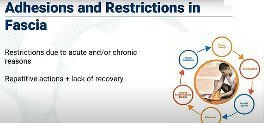
Adhesions and restrictions in fascia
adhesions and restrictions in fascia occur due to acute and or chronic reasons
Body injury due to repetitive actions or stress often combined with a lack of adequate recovery
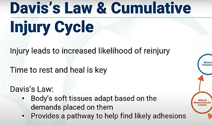
DAVIS’s LAW and CUMULATIVE INJURY CYCLE
injury leads to increased likelihood of re-injury
Taking time to rest and heal is the key
Davis law:
Body’s soft tissues adapt based on the demands placed on them
Provides a pathway to help find likely adhesions
FITT
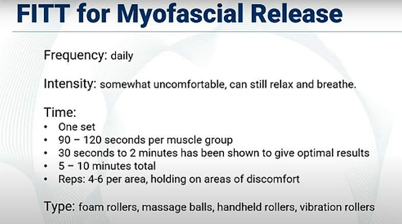
FREQUENCY - Daily
NTENSITY - Somewhat uncomfortable, can still relax and breathe
TIME
- 1 set
- 90-120 seconds per muscle group,
- 30 sec to 2 min - optimal results
- 5-10 min total
- 4-6 active rolls per area, holding on areas of discomfort
TYPE - Foam rollers, massage balls, handheld rollers, vibration rollers
MED
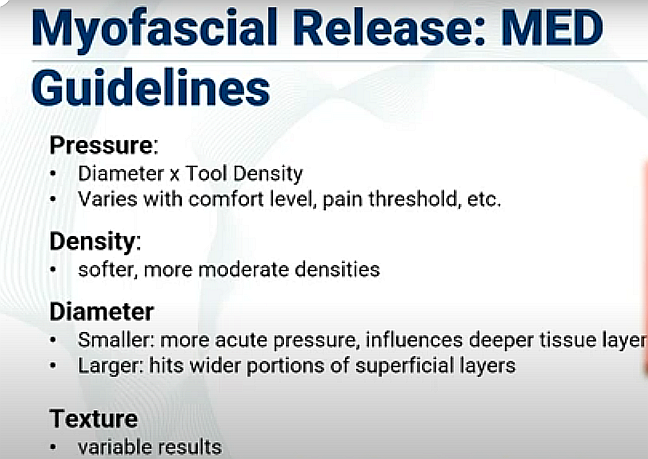
Pressure
Diameter x Tool density
Varies with comfort level, pain threshold, etc.
Density:
softer = more moderate densities
Diameter:
smaller diameter gives more acute pressure, influences deeper tissue layer
Larger diameter hits wider portions of superficial layers
Texture:
variable results
Manual Therapy Tools
Stick-handheld device that can be used like a rolling pin. Best for lower legs especially the calves
Vibration therapy tool- deliver rapid high frequency vibrations to the affected area, which can help to break up adhesions that are causing pain and discomfort. Come in a variety of forms, from handheld versions to larger devices like massage guns, and can be used on various parts of the body, including the back, legs, and arms.
Hands
Massage technique that involves applying sustained pressure to specific areas of tightness or discomfort in the muscles and fascia. Gentle pressure is applied with fingers, palms, or even elbows along the affected area. Using a stretching or pulling movement is encouraged to release tension. Can be highly effective way to address muscle tension and promote overall relaxation and wellness.
Electrotherapy
Involves the use of electrical stimulation to target areas of muscle tension and tightness, electrodes are placed on the skin to deliver electrical impulses to the affected muscles and fascia. Help to reduce muscle spasms, increase blood flow, and encourage muscle relaxation. Stimulate the release of endorphins, the body’s natural pain relievers, promoting a sense of well-being.
Gua Sha
Offer a soothing touch to tight and knotted muscles, helping to release stored tension and promote a sense of relaxation. Can sculpt and smooth out areas of discomfort, allowing the body's natural healing processes to begin
Facial roller
Skincare tool used for facial massage and skincare routines. Typically consists of a roller with one or two smooth, rounded heads made of materials such as jade, rose quartz, or stainless steel. Designed to gently roll over the skin, providing a cooling, soothing effect. Believed to offer multiple benefits for the skin; can help improve blood circulation, decrease puffiness, and promote lymphatic drainage
Rollers and balls
Offer a pathway to address tightness, tension and restrictions within fascia. Help release adhesions, improve blood flow, enhance the flexibility and mobility of the body. Allow the fascia to soften and regain its natural elasticity. Promotes physical healing, a deep sense of connection and mindfulness.
Cupping
Specialized cups are placed on the skin to create a suction effect. Targets and stretches the fascia. Pulling action helps to alleviate tightness, reduce pain, and improve circulation, allowing the body to heal more effectively, release adhesions and promote the flow of energy.
Peanut
Double-ball tool designed to apply pressure in a specific area, allowing for concentrated release of tension and adhesions within fascia. Placed between the muscles or along the spine and rolled, it compresses and releases to alleviate tightness, improve circulation, and stimulate the nervous system
QUIZ
Identify which component of the FITT principle each example applies to
Daily - FREQUENCY
Somewhat uncomfortable, can still relax and breathe - INTENSITY
1 set, 90-120 seconds per muscle group, 4-6 active rolls per area, holding on areas of discomfort - TIME
Foam rollers, massage balls, handheld rollers, vibration rollers -TYPE
PRECAUTIONS and CONTRAINDICATIONS
PRECAUTIONS:
Local tissue inflammation
Hypertension
Diabetes
Varicose veins
Scoliosis
Fibromyalgia
Recent injuries/surgeries
CONTRAINDICATIONS:
Open wounds
Pregnancy
Bone fractures
Deep vein thrombosis
Myositis ossificans
Osteomyelitis
Osteoporosis
Bleeding disorders
Cancer or malignancy
Connective tissue disorders
Screen for precautions and contraindications prior to using any MRT on your own, or with others.
It is important to check with a health care professional.
TERMINOLOGY
Myofascial Release Therapy
Massage treatment-based therapy where the focus is to create change in the myofascial component of the tissue, restoring mobility and tissue tolerance
Muscle Spindles
Stretch receptors that run parallel to muscle fibers that send information on changes in muscle length
Minimum Effective Dosage (MED)
The minimum effective dose of an input required to receive a desired outcome
Golgi Tendon Organs
Sensory mechanoreceptors located near muscle tendons
Davis’s Law
The body’s tissues adapt based on the demands placed upon them
Cumulative Injury Cycle
When an injury occurs, if it doesn’t receive enough time to recover then it is more likely to be injured again
Autogenic Inhibition
Golgi tendon organs sense the development of high tension and send a signal to the muscle to relax
